Cryo-Electron Tomography

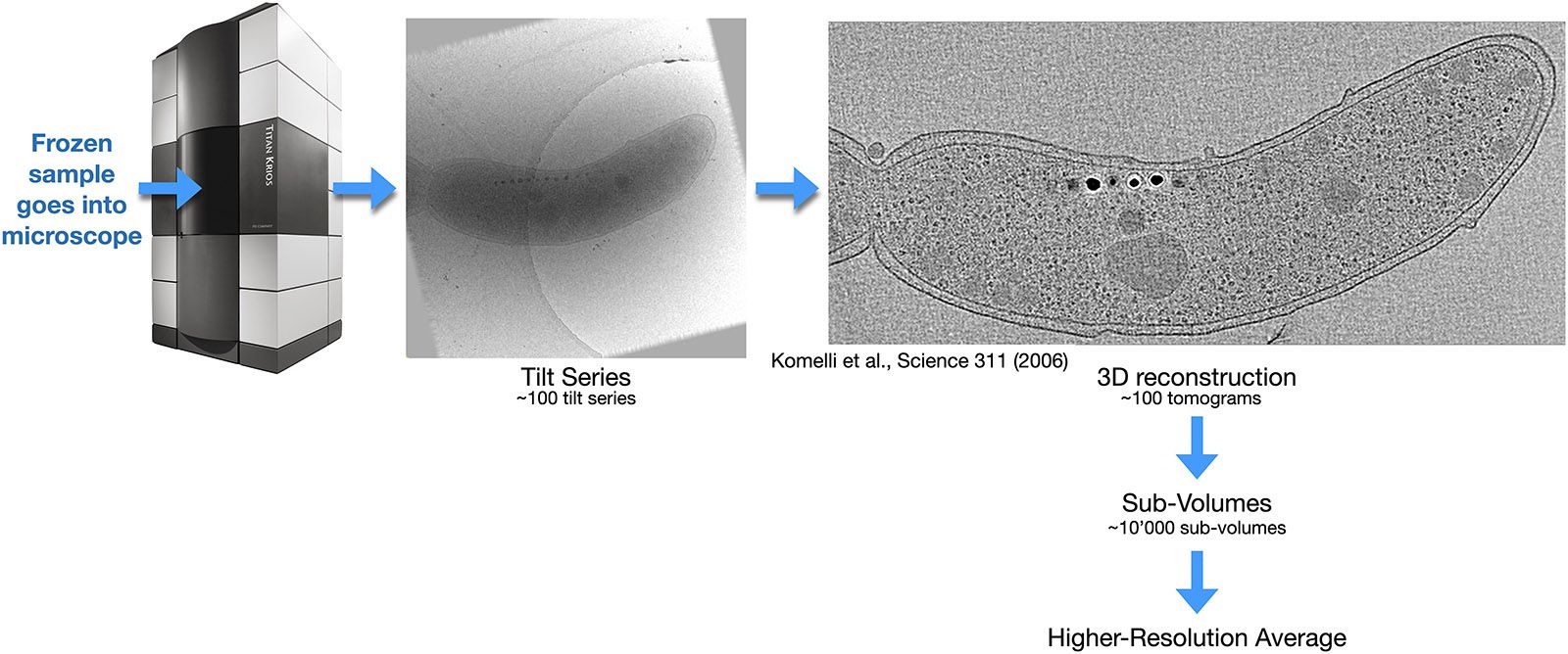
In electron tomography, each object of interest in the sample is imaged horizontally and at various angles to the electron beam. The recorded series of images from different tilt angles is then called a "tilt series". The increase in thickness to the electron beam of the tilted sample limits the possible angular range to ±60°. Computer image processing of the images gives a 3D reconstruction of the sample, yet a lower resolution, because individual movements of sub-sections of the sample during recording of the tilt series cannot be compensated at this step (it will be compensated later, see below). A first 3D reconstruction from a tilt series is therefore often limited to 2nm 3D resolution. Because of the limited tilt range, the whole 3D Fourier space cannot be sampled, so that the resolution in the z direction is lower than in the X and Y directions.

Recorded tilt series and their first 3D reconstruction can then be inspected manually or by computer software, to identify regions of interest. If for example the several ribosomes in a cell would be the objects of interest, then the X/Y/Y coordinates of each ribosome could be detected. If software then extracts from each two-dimensional image from the tilt series the image segment that belongs to a certain ribosome, then for each ribosome the computer would obtain a set of individual images at different tilt angles. These could then be further processed in a single-particle-like approach, which allows movie-mode drift correction and precise correction of electron microscopy artifacts (such as the CTF), so that a much higher resolution can eventually be achieved at the end of this process. In several cases, this single-particle processing of electron tomography tilt series led to side-chain-resolving resolution on proteins that were still within the cellular enviroment.

Sample Requirements for Cryo-Electron Tomography+-

Cryo-ET requires the sample to be 300nm thin or thinner. Biological cells or tissue that is thicker needs to be vitrified (i.e., frozen to LN2 temperature without ice crystal formation), and then sectioned to thin lamellae. This can be done with a cryo-ultramicrotome or with a cryo-FIB-SEM, both of which are available at the EMF at UNIL.

The work flows are vast and very complex for sample handling, fixation or staining of fluorescent labeling, freezing, sectioning, and correlative imaging with a light microscope or other instruments prior to the cryo-ET investigation. Please contact us for discussing your project and identifying a suitable sample handling strategy.

Once suitably thin cryo-preparations of the biological sample are available, these cryo-EM grids can then be imaged in a 300kV Titan Krios at LN2 temperature in so-called tilt series. During this procedure, the sample is imaged as dose-fractionated movies in the Titan, while the sample is rotated to various different tilt angles. This results in a series of images, each recorded at a different sample tilt angle. This "tilt series" of images can then with computer image processing be transformed into a first 3D reconstruction of the sample. 

Sub-volumes containing similar proteins from the cell (e.g., lots of ribozomes) can then be localized in this 3D reconstruction with computer help, and a larger number of such extracted sub-volumes can then be analyzed, classified, aligned and averaged, to yield a high-resolution representative structure of that molecule within the cellular context. Such sub-volume analysis (SVA) can in favorable cases sometimes reach very high resolution, so that even secondary structure elements or still higher resolution can be recognized from a sample that was never purifed. 

Extensions of Electron Tomography+-

Electron tomography can be used to :

Extend the information obtained by Serial Block-face Imaging in the scanning electron microscope.

When areas of interest are located by room temperature serial block-face imaging, the resin-embedded sample (e.g., small region of tissue or a small region of a mouse brain) can be removed from the microscope (SEM), and mounted in a conventional ultramicrotome, where thicker (e.g. 100 to 400 nm) sections are cut. These sections are then sequentially mounted on electron microscopy grids and analyzed by electron tomography in a transmission EM, e.g., the FEI Titan. Together the images reveal the 3-D structure of the resin-embedded sample at a resolution of about 30 nm.  

Examine cryo-sections of samples that were embedded in resin at cryo temperatures. Sub-nanometer resolution can be achieved. 

• Examine cryo-sections of samples embedded in vitreous ice (CEMOVIS). The 3D structure of the sample under close-to-native conditions is obtained at nanometer resolution.

Cryo-ET image processing+-

Recorded data will be computer processed during data collection on DCI computers, so that a first 3D reconstruction is available during data collection. Subsequent in-depth processing of recorded images requires computing hardware with GPUs that have generous RAM on each GPU card, certain software systems, and expertise, all of which is available at the DCI. Please contact us for more info.